top of page
MAYA DEZA CULBERTSON SCOTT
RESEARCH BIOLOGIST
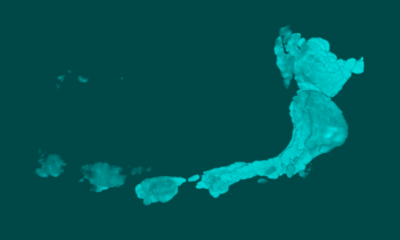
 Zebrafish embryo (Danio rerio)Z-projection multichannel confocal image of a 4-day post-fertilization zebrafish embryo. The developing, cartilaginous cranial skeleton is visualized with Collagen-IIa immunostaining (white). NeuroD:GFP transgenic expression is represented by a pseudocolored heat map (magenta-violet-orange). Nuclei in all tissues are counterstained with DAPI (blue). |  Cranial Ganglia, phox2bExtended focus light-level image of the region around the otic vesicle in a 2-day post-fertilization zebrafish embryo. The developing cranial ganglia are visualized via NBT-BCIP in situ hybridization for the phox2b transcript (purple). Image modified from Figure 2, Culbertson, et al., 2011 (PLoS ONE). |
|---|---|
 3D Reconstruction, Cranial Ganglia3D reconstruction from a high-resolution confocal z-stack of the cranial ganglia in a 4-day post-fertilization zebrafish embryo immunostained for the neural marker Elavl 3/4 (or Hu C/D). | 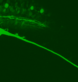 Axon OutgrowthDetail from time lapse image showing the outgrowth of collateral axons over ~17 hours. Images obtained over 15 minute intervals. |
 Tissue Section In-Situ, foxd3Transverse 20-micron frozen section of an 18-hour post-fertilization zebrafish embryo showing neural crest derived glial precursor cells via NBT/BCIP in-situ hybridization for foxd3 (purple). Image modified from Figure 1, Culbertson, et al., 2011, PLoS ONE). |
© 2014 by Maya Deza Culbertson
bottom of page